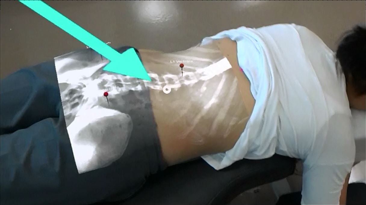
 March 11, 2019 – A recently published University of Alberta study is looking at utilising Microsoft HoloLens technology to reunite patients with their X-ray images.
Greg Kawchuk, a professor in the Department of Physical Therapy, Faculty of Rehabilitation Medicine at the University of Alberta, has been working with Department of Computing Science professor Pierre Boulanger and researchers from the University of Southern Denmark to make ‘X-ray vision’ a reality.
“I’ve been following this technology from day one.” Said Kawchuk. “I knew right away that it could be used to help health-care professionals, such as physicians, physiotherapists and chiropractors, locate anatomy that was not apparent to normal vision. [My team and I] bought the goggles as soon as they hit the market.”
Kawchuk and his team chose to look at Microsoft Hololens for this specific study. With HoloLens, the team is able to project a holographic, mixed-reality superimposition of a person’s X-ray onto their back. Physicians wearing the headset will be able to see an anatomically correct view of the person’s spine. This helps with adjusting treatment and exercise plans.
“Traditionally, educators had to point to where organs were located in the body, or draw images on people’s skin so they can imagine where body parts are located,” added Kawchuk. “Now we don’t need to do that. We can just put on a pair of goggles and ‘see’ what’s underneath, like true X-ray vision.”
Published in February in PeerJ, the study conducted by Kawchuk and his team found that HoloLens was 73 percent accurate when it came to locating vertebrae levels in the spine. A total of 13 participants who already had pre-existing spine X-rays took part in the study.
According to Kawchuk, there are positives for both the patients and the clinicians, but it really all boils down to having the ability to include the patient in their own data:
“The historic disconnect between X-ray and patient is the basis of the well-worn clinical advice to ‘treat the patient, not the film.’ Disconnected imaging can result in misguided interpretations of the image with respect to the patient.”
While the technology will make an impact in the clinical community, there’s also room for the tool to be used in an educational setting.
“This is perfect for classroom education. Not just for healthcare providers and their patients, but also for students who are going to be entering into the field,” said Kawchuk.
“Future plans are already in the works to take the new technology into a clinical environment to see how it can improve interactions between physicians and patients when it comes to explaining imaging results.”
Image credit: University of Alberta / Greg Kawchuk / Rehab Robotics Lab
About the author
Sam is the Founder and Managing Editor of Auganix. With a background in research and report writing, he has been covering XR industry news for the past seven years.
